

Advanced Brain Tumor Resection Techniques: The International Standard for Maximal Safe Removal

Receiving a brain tumor diagnosis is life-changing, but advancements in neurosurgery have fundamentally transformed the treatment landscape. The primary goal of modern brain tumor surgery is no longer merely survival, but achieving maximal safe resection (MSR)—removing the largest possible volume of the tumor while rigorously preserving the patient’s neurological function and quality of life. This paradigm shift requires integrating sophisticated, real-time technologies that allow neurosurgeons to see, navigate, and operate with unparalleled precision.
This evolution is driven by compelling evidence: a greater extent of resection (EOR), especially in high-grade gliomas like Glioblastoma (GBM), directly correlates with improved progression-free and overall survival rates. For international patients seeking the best neurosurgeon in India, understanding these advanced techniques is crucial to choosing a center that meets global standards of care.
The Goal: Maximal Safe Resection (MSR)
MSR is the core philosophy guiding elite neurosurgical teams worldwide. Unlike historical approaches, which relied heavily on pre-operative scans and visual estimation, MSR uses cutting-edge tools to achieve two critical outcomes simultaneously:
- Maximizing Tumor Removal: Aggressively targeting the tumor mass, including microscopic cells extending into seemingly healthy tissue, which improves prognosis.
- Minimizing Neurological Deficit: Protecting eloquent brain areas (those responsible for speech, movement, sensation, and memory) to ensure the patient retains their cognitive and functional independence.
Achieving this balance is only possible through the synergistic use of advanced intraoperative technologies.
Foundational Technologies for Precision Neurosurgery
These are the non-negotiable tools that form the backbone of a high-volume, international-standard neurosurgical practice:
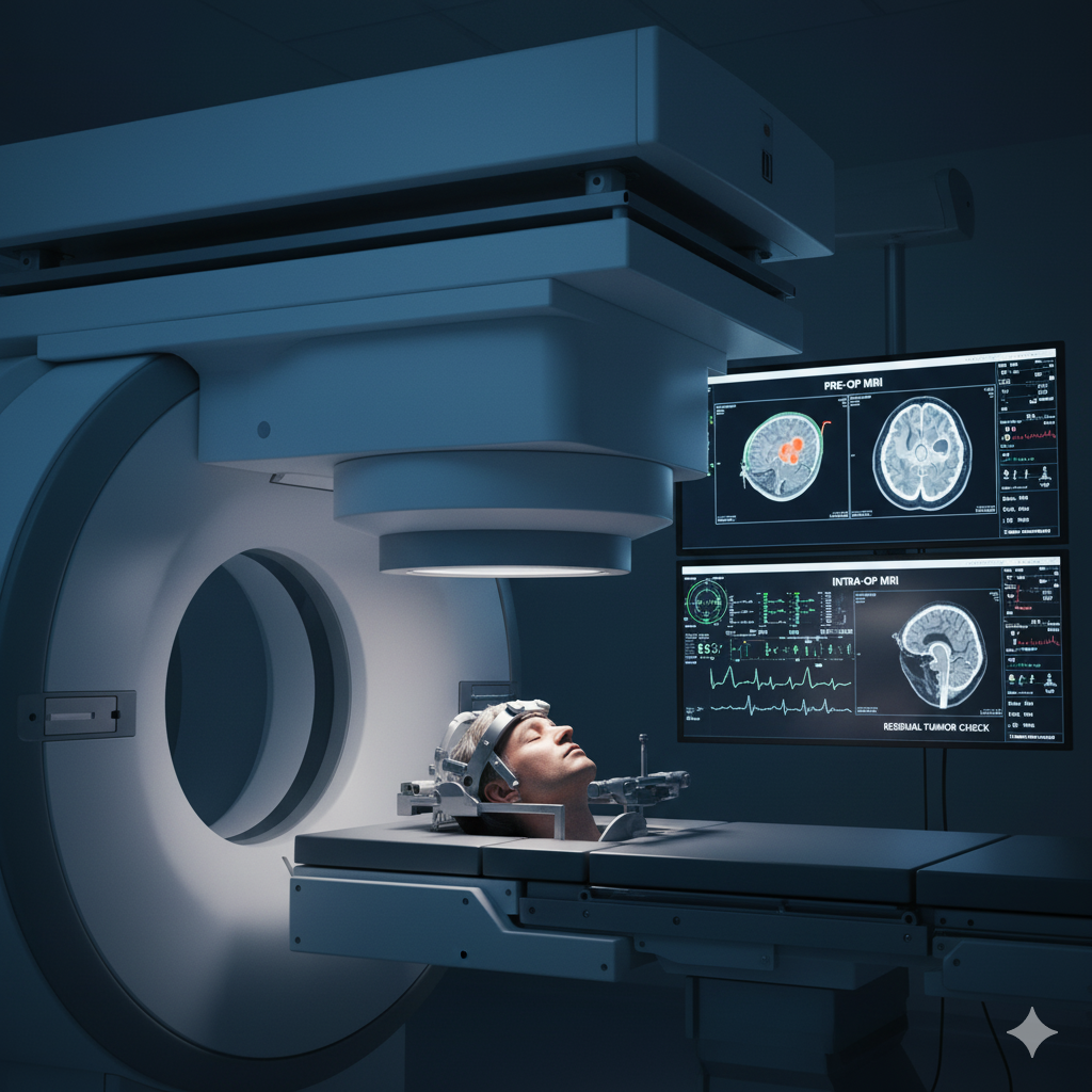
1. Intraoperative Magnetic Resonance Imaging (iMRI)
What it is: iMRI integrates a high-field MRI scanner directly into the operating room, allowing the surgeon to obtain updated, high-resolution scans of the patient’s brain during the surgery without moving the patient.
2. Fluorescence-Guided Surgery (FGS) with 5-ALA
What it is: FGS utilizes a special oral compound, 5-aminolevulinic acid (5-ALA), given to the patient before surgery. Once metabolized, the compound causes malignant tumor cells (gliomas) to fluoresce or glow a vivid pink-red under a specialized blue-light microscope, while healthy brain tissue appears blue.
Why it is a game-changer:
- Sharp Margin Delineation: The difference between a high-grade tumor and normal brain is often invisible to the naked eye. 5-ALA provides a biological contrast, allowing the surgeon to identify the exact tumor margins, including subtle infiltrating areas.
- Proven Survival Benefit: International multi-center trials have confirmed that using 5-ALA-guided resection improves the rate of GTR and, critically, extends the progression-free survival for patients with malignant gliomas. This is a vital technique utilized by the best neurosurgeon for brain hemorrhage and tumors alike.
3. Advanced Neuronavigation and Tractography
What it is: A GPS system for the brain. Neuronavigation uses pre-operative imaging (MRI, CT) merged with advanced imaging techniques like Diffusion Tensor Imaging (DTI) to create a precise, 3D map of the brain. DTI specifically maps the white matter fiber tracts (the brain’s highways) responsible for critical functions.
How it ensures safety:
- Pre-Surgical Planning: Allows the surgeon to map the safest path to the tumor, bypassing critical motor, sensory, and language pathways.
- Real-Time Guidance: The neurosurgeon’s instruments are tracked on the 3D map during the procedure, providing continuous, sub-millimeter guidance that prevents accidental injury to functional tracts.
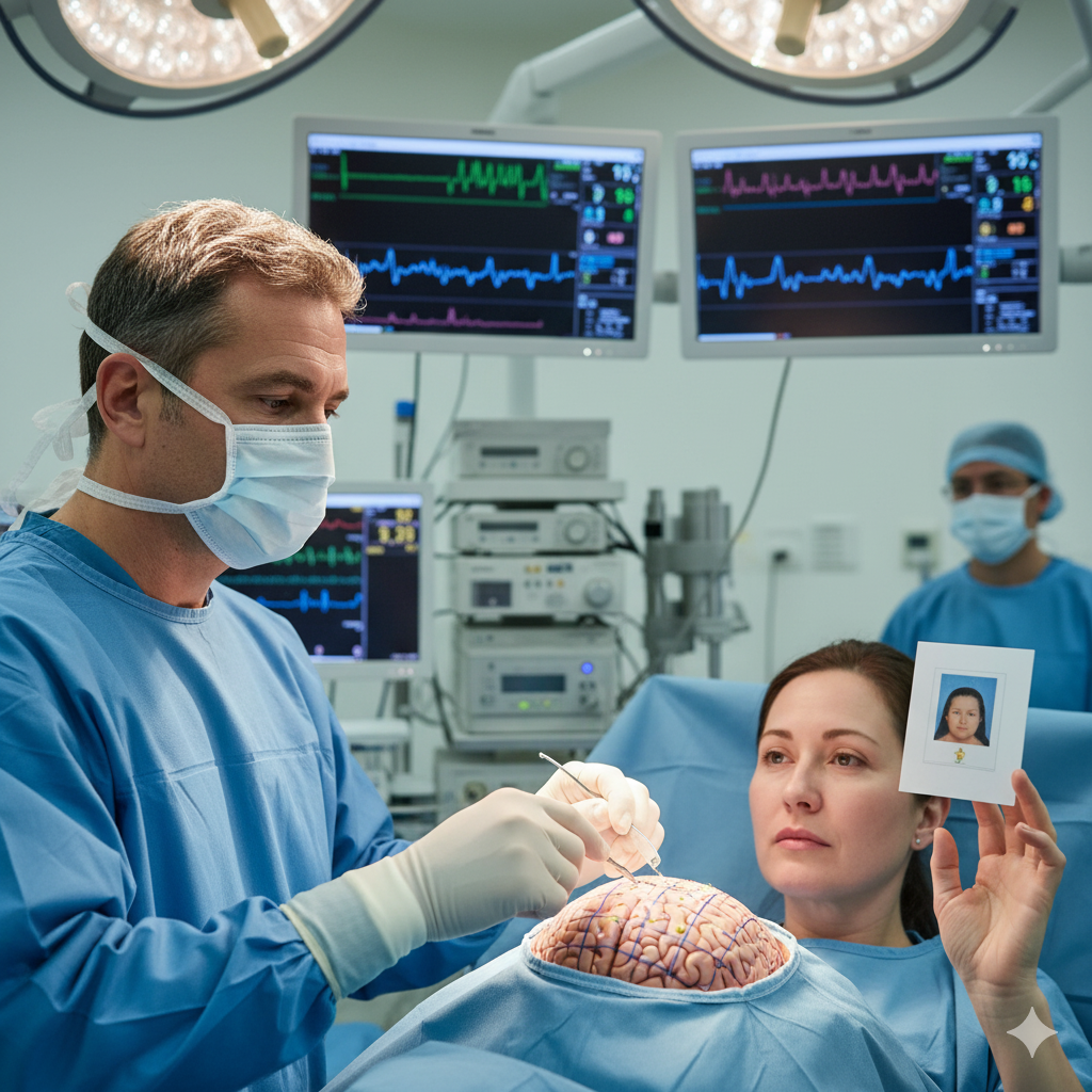
The Apex of Functional Preservation: Awake Craniotomy
For tumors located in or immediately adjacent to eloquent areas (brain regions controlling movement or language), the Awake Craniotomy technique is the gold standard for prioritizing long-term quality of life.
The Procedure in Detail:
- Patient Preparation: The patient is sedated for the initial phases of the procedure (opening and closing the skull bone).
- Mapping and Stimulation: Once the tumor is exposed, the patient is gently awakened. The neurosurgeon uses a mild electrical current (Direct Electrical Stimulation, or DES) to map the brain’s surface in real-time.
- Real-Time Monitoring: As the surgeon removes the tumor, the patient performs tasks (e.g., naming objects, moving limbs) under the supervision of a specialized team (neurophysiologists, anesthetists).
- Preservation Guarantee: If stimulation causes a temporary disruption of a function (like slurring of speech), the surgeon immediately knows that area must be preserved, ensuring that the best neurosurgeon for DBS and functional procedures can achieve maximal resection without causing permanent deficit.
This technique transforms a potentially disabling surgery into a function-preserving one, offering patients the best neurosurgeon in India experience by aligning oncological success with functional recovery.
Emerging Technologies Driving the Future of Resection
The global push towards less invasive and more targeted surgery continues with these innovations:
- Laser Interstitial Thermal Therapy (LITT): A minimally invasive, catheter-based technique where a laser fiber is inserted into the tumor through a small burr hole. The laser heats and destroys tumor tissue under continuous, real-time iMRI thermal monitoring. LITT is an excellent option for deep-seated or recurrent tumors that are difficult to reach conventionally.
- Exoscopic and Robotic Assistance: High-definition 3D visualization systems (exoscopes) provide a magnified, brilliant view of the surgical field, which is often superior to traditional microscopes. Robotic platforms offer enhanced dexterity, tremor reduction, and stable visualization, further increasing the precision available to the top neurosurgeon in India.
Why International Patients Choose Centers Using Advanced Techniques
Patients traveling internationally for neurosurgical care often have complex, challenging tumors that require this high level of technological expertise. When choosing a specialist like Dr. Aditya Gupta, patients are seeking a neurosurgeon who:
- Has Verified Experience: Regularly performs complex tumor resections using combined modalities (iMRI + 5-ALA + Neuronavigation).
- Prioritizes Function: Views the surgical outcome through the lens of functional preservation (using Awake Craniotomy when needed).
- Adheres to Global Protocols: Follows the latest international guidelines (like those from the World Health Organization and leading neuro-oncology societies) for maximal safe resection in all types of brain tumors, from gliomas to meningiomas.
Conclusion: Precision and Expertise Define Modern Neurosurgery
The treatment of brain tumors today is an intricate synergy of expert surgical hands and state-of-the-art technology. The commitment to Maximal Safe Resection—driven by tools like iMRI, 5-ALA Fluorescence, and Awake Mapping—provides the best chance for both long-term survival and preservation of a high quality of life.
If you or a loved one are facing a complex brain tumor diagnosis and are seeking a world-class neurosurgical opinion that meets the highest standards of international care, please contact our office to discuss a personalized treatment plan with a leading expert. Book an appointment today to learn how advanced techniques can redefine your prognosis.
(Glioblastoma Treatment Guidelines): Glioblastoma is the most aggressive form of brain cancer, making maximal safe resection crucial. Read more on the impact of extent of resection from the National Institutes of Health. https://www.ncbi.nlm.nih.gov/
( 5-ALA Fluorescence in Surgery): The use of 5-Aminolevulinic acid (5-ALA) is a major advancement in visualizing malignant tumors. Understand its clinical efficacy as detailed by the European Association of Neuro-Oncology. https://www.eano.eu/ (Using EANO as a representation for a global guideline body, actual link will be to a relevant high-authority study on 5-ALA).
(Overview of Minimally Invasive Neurosurgery): Minimally invasive techniques, including LITT and keyhole surgery, are revolutionizing recovery. Explore these techniques at the Mayo Clinic. https://www.mayoclinic.org/
- Tags:
- 5-ALA fluorescence
- awake brain surgery
- Awake Craniotomy
- best neurosurgeon for brain hemorrhage
- best neurosurgeon for brain tumor
- best neurosurgeon for DBS
- best neurosurgeon in India
- brain shift correction
- brain tumor resection techniques
- brain tumor surgery techniques
- eloquent area tumor
- Fluorescence-Guided Surgery (FGS)
- functional preservation neurosurgery
- glioblastoma surgery
- GTR brain tumor
- high-grade glioma treatment
- iMRI for brain tumor
- Intraoperative MRI (iMRI)
- laser interstitial thermal therapy
- LITT for brain tumor
- maximal safe removal
- Maximal Safe Resection (MSR)
- neuro-oncology treatment
- neuronavigation surgery
- neurosurgery technology
- neurosurgical innovations
- skull base tumor removal
- Top neurosurgeon in India